

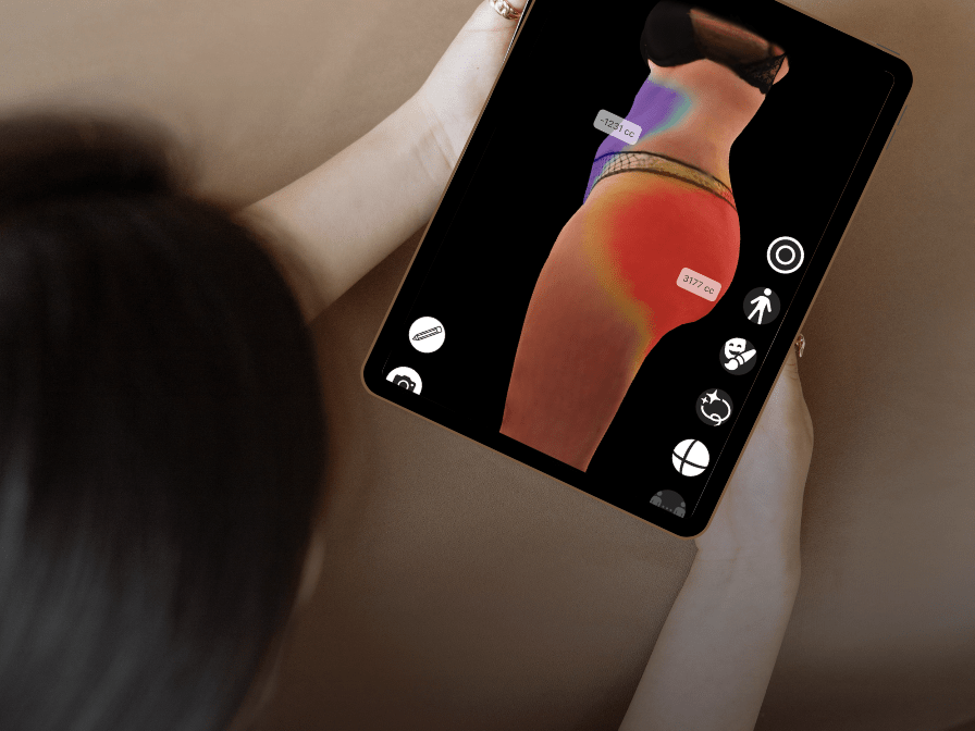
Today, we want to talk about something that rarely enters the conversation around aesthetic medicine: the science behind modern body visualization.
We sat down with Endri Dibra, CEO of Arbrea Labs, whose PhD research at ETH Zurich on 3D body reconstruction using deep learning became one of the early foundations for how surgeons and patients visualize body transformation today.
His research papers, HS-Nets: Estimating Human Body Shape from Silhouettes with Convolutional Neural Networks and Human Shape from Silhouettes Using Generative HKS Descriptors and Cross-Modal Neural Networks, introduced one of the first deep learning systems capable of reconstructing a full 3D human body from only two photographs, years before generative AI and neural rendering became mainstream topics.
Interestingly, the relevance of this work has only increased over time.
When Endri published this research in 2016, body visualization was still largely experimental. Today, the medical landscape has changed dramatically. The rise of GLP-1 medications such as Ozempic and Wegovy is creating rapid body transformations that patients increasingly want to understand visually, not just numerically. Conditions such as Lipedema are finally entering mainstream clinical discussion, creating demand for better tools to map, track, and visualize the body over time.
Beyond surgery itself, the implications extend into recovery monitoring, compression garments, digital consultations, and patient confidence throughout the treatment journey. What started as research into reconstructing the human body from silhouettes is now becoming part of the infrastructure for how medicine understands the human form digitally.
You can also read Endri’s reflections on the research and its long-term impact in his recent LinkedIn post.
”Arbrea. Never. Stops.
Dr. Endri DibraArbrea Labs

Interview
The paper introduced one of the first deep learning systems to reconstruct a full 3D body from just two photographs. Why was two views the key technical decision?
At the time, most 3D reconstruction systems relied on expensive scanners, many synchronized cameras, or highly controlled environments. That made the technology difficult to scale clinically or use in everyday settings. The key insight was that the human body already contains a huge amount of geometric information in its silhouette. A front and side view together constrain the body shape surprisingly well. By combining those two views with statistical body models and deep learning, we could estimate a realistic 3D body without requiring specialized hardware.
Two images were also a practical decision. If you ask patients to capture ten images or use external sensors, adoption drops immediately. But two smartphone photos are simple, fast, and globally scalable. That principle still drives many of the design decisions we make today at Arbrea.
How does that translate into what Arbrea Body does today?
The core philosophy remains the same: making advanced body visualization accessible in real clinical workflows.
At Arbrea Labs, we evolved those early research concepts into a real-time platform for aesthetic and reconstructive consultations. Instead of only reconstructing body geometry, Arbrea Body combines computer vision, AI, AR, and simulation technology to help surgeons and patients communicate visually during consultations. The important part is not only realism. It is trust and communication.
Visualization creates a shared language between surgeon and patient. The technology helps reduce uncertainty while keeping the surgeon at the center of the consultation process. What started as academic research into silhouettes became a clinical communication tool.
Where do you see this going next? What’s the problem that still needs solving?
Today, most systems focus on a single moment in time: one consultation, one scan, one simulation. But the human body changes continuously due to aging, surgery, hormones, weight changes, pregnancy, medications like GLP-1s, recovery processes, or medical conditions such as lipedema. The real challenge is building systems that can understand and visualize those changes over time responsibly and accurately. That requires more than computer graphics. It involves biomechanics, AI, privacy-preserving computation, medical validation, and patient-centered design. It also means moving from static visualization toward personalized predictive modeling.
The body is dynamic. Digital medicine still largely treats it as static.
The paper showed your system ran in under half a second per image, orders of magnitude faster than anything else at the time. Why did speed matter so much?
Speed was essential because interaction changes behavior.
If a system takes minutes to reconstruct a body, it becomes an offline technical process. But if results appear almost instantly, it becomes part of a natural conversation between doctor and patient. Real-time feedback creates engagement. A surgeon can adjust inputs, explain proportions, discuss options, and iterate collaboratively during the consultation itself.
That was one of the early realizations that later became central to Arbrea’s products: latency is not only a technical metric. It fundamentally changes user experience and adoption. Even today, achieving high-quality simulation directly on mobile devices, without sending patient data to the cloud, remains one of our biggest engineering priorities.
Your paper reported a measurement error of around 4mm across 16 body measurements. How important is that level of precision when you’re talking about surgical planning rather than garment fitting?
Precision matters, but context matters even more.
Human bodies are naturally variable. Even clinical measurements taken manually by different professionals can vary by several millimeters. So achieving an average error around 4mm from only two RGB images was a significant step at the time, especially given the speed and accessibility of the system.
However, surgical planning is not only about absolute measurement precision. It is also about proportionality, anatomical consistency, communication, and expectation management.
The goal was never to replace surgical judgment. The goal was to augment communication with data-driven visualization.
What is Arbrea Body?
Arbrea Labs develops AI-powered visualization tools for aesthetic and reconstructive medicine.
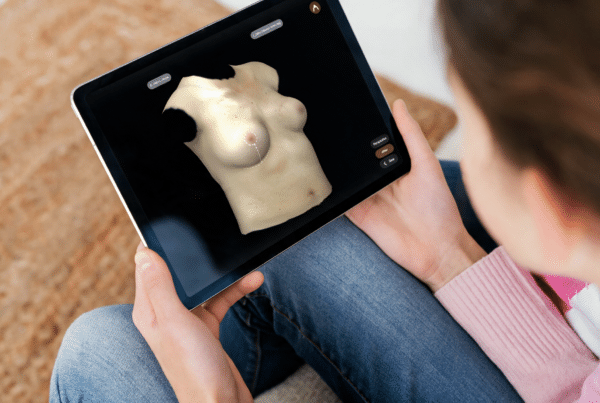
Arbrea Body enables surgeons to perform real-time body simulations directly on mobile devices and tablets without requiring external scanners or cloud-based processing. The platform is designed to support consultations, patient communication, treatment planning, and longitudinal visualization throughout the patient journey. Built on years of research in computer vision, deep learning, and augmented reality, the technology combines privacy-focused on-device AI with clinically oriented workflows.
The broader vision is to create a unified digital ecosystem connecting visualization, consultation, follow-up, and patient engagement across the entire aesthetic and reconstructive journey.
Reference
[1] Dibra, Endri, et al. “Hs-nets: Estimating human body shape from silhouettes with convolutional neural networks.” 2016 fourth international conference on 3D vision (3DV). IEEE, 2016.






 English
  Portuguese
  Italian
  Spanish
  German
  Arabic
  French
  Korean
  Albanian
  Thai
Thai  Hindi
Hindi  Vietnamese
  Japanese