

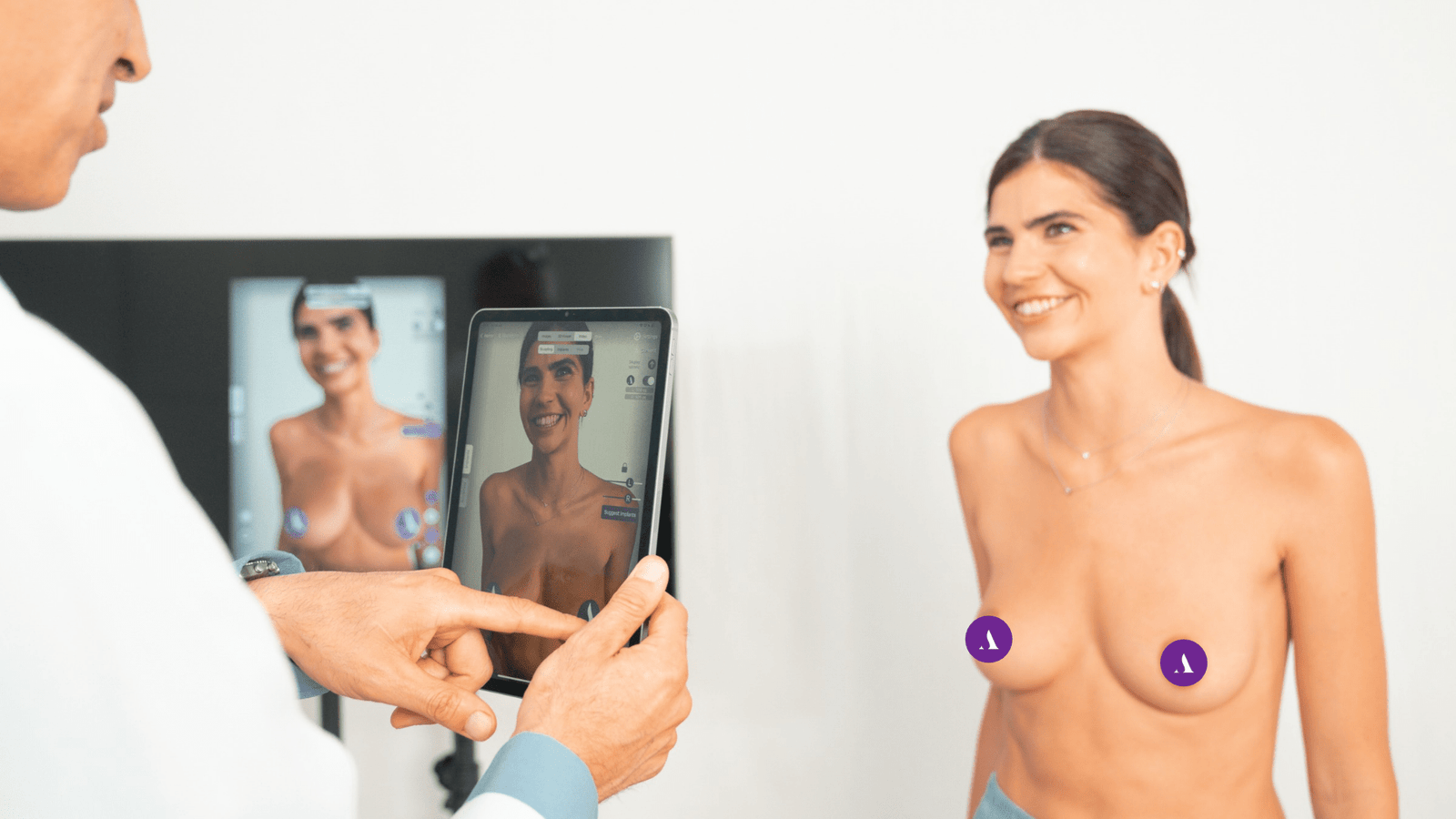
A patient comes back for their six-month follow-up. The result looks good genuinely good. But six months ago, the simulation you planned around projected something slightly different: a bit more projection here, a slightly different contour there. Was the outcome actually close to the prediction, or did it just look close enough in a photo that nobody checked?
That question is harder to answer than it should be, and the reason comes down to a tool most practices still rely on by default: the photograph. A simulation can look flawless and still be wrong. That’s the uncomfortable truth behind predictive modeling in aesthetic medicine: a rendered outcome can be visually convincing while sitting several millimeters off from what the patient actually ends up with. This is exactly why LiDAR 3D scanning accuracy in plastic surgery has become such an important question over the past few years, because a photograph was never built to measure anything. It was built to be looked at.
This is the problem depth-sensing technology was designed to solve, and it’s why smartphone LiDAR scanning has quietly become one of the more consequential additions to outcome documentation in aesthetic practice.
3D scan vs photo surgical outcome: why the difference matters
Every photo is a two-dimensional projection of a three-dimensional object, captured under one specific lighting condition, from one specific angle, at one specific moment. Move the light, tilt the camera two degrees, or catch the patient mid-breath, and the same anatomy can look meaningfully different. None of that variation is real change, it’s noise. But if a photo is your only outcome record, there’s no way to separate the noise from the signal. That’s the core of the 3D scan vs photo surgical outcome debate: a photo shows you a result, a scan measures it.
LiDAR (Light Detection and Ranging) removes that ambiguity by measuring geometry directly. Rather than inferring shape from light and shadow, the sensor emits thousands of infrared pulses per scan and times how long each one takes to return, a technique called time-of-flight ranging. The result is a literal map of the surface: real coordinates, real distances, real curvature. Put simply: a photo tells you the result looked right. A scan tells you exactly how right, down to the millimeter.
How accurate is smartphone LiDAR scanning for surgery?
This isn’t a claim taken on faith. Over the past several years, a specific body of research has directly tested smartphone LiDAR scanning accuracy against dedicated clinical-grade 3D imaging systems, and the results have been consistent:
- A 2024 clinical study testing LiDAR-based scanning apps on the iPhone 14 Pro against a stationary clinical imaging reference found mean surface deviations of just 1.0–2.0 mm across facial landmarks, well inside what’s considered clinically usable for surgical planning.
- A separate 2025 study on breast measurements using the iPhone 15 Pro’s LiDAR sensor compared it against manual tape measurements in 25 patients undergoing breast procedures, finding very good accuracy for sternal notch-to-nipple and nipple-to-midline distances, with the one weaker spot being the nipple-to-inframammary fold measurement — a single anatomically tricky landmark, not a failure of the scan geometry as a whole.
- One of the most recent validation studies, published in May 2025, tested a smartphone LiDAR/TrueDepth-based scanning app against CBCT imaging in 30 patients and found trueness values of 0.70–0.85 mm — still comfortably within the sub-1mm range considered clinically reliable.
Taken together, these studies answer the accuracy question directly: consumer-grade LiDAR, in the hands of a clinician, produces measurement-grade data, at a fraction of the cost and setup complexity of dedicated scanning hardware.
In short: the device already in your pocket can now measure a surgical result almost as precisely as equipment that costs tens of thousands of dollars.
Predictive simulation accuracy in plastic surgery: why measurement matters
Here’s where it becomes more than an interesting sensor spec. A simulation is a prediction, and predictive simulation accuracy in plastic surgery is only as credible as the evidence used to check it. If the only thing you can compare a predicted outcome against is a photo, you can say the result “looked similar” — but you can’t say by how much it was off, or where, or why.
A post-treatment LiDAR scan changes that. It can be aligned directly against the pre-treatment simulation, point cloud to point cloud, producing an actual number: this many millimeters of deviation, at this specific location. That’s not a subjective impression, it’s a measurement. And a model corrected against real, quantified error gets sharper in a way that visual comparison never allows.
This is precisely why, inside the Research Partner Program, live case contributions require a LiDAR-capable device. An archive case can be built from documentation a doctor already has. A live case exists specifically to close the loop, simulation against reality, measured, not estimated. Every scan submitted this way becomes a data point tied to a real technique, a real implant or product choice, a real patient population — the exact material that narrows the distance between what Arbrea predicts and what a doctor actually delivers, across Arbrea Breast, Arbrea Face, and Arbrea Body alike.
Why this program exists
Simulation quality is capped by the reality it has seen. A model trained only on generic, anonymized datasets can produce a plausible-looking prediction, but plausible isn’t the same as accurate to your technique, your implant or product choices, or your patient population.
That’s the entire premise behind the Research Partner Program: a limited circle of doctors worldwide contributing real, measured cases from their own practice, so the model improves against reality instead of assumption. Every case sharpens fitting, prediction, and scan interpretation a little further, and every improvement ships back into the Suite you already use. It’s a direct exchange: your documented work makes the model better, and a better model gives you back more accurate simulations to plan and bonus.
The takeaway
A photo can tell you a result looked good. Only a measurement can tell you how close it actually came to plan. LiDAR scanning makes that measurement possible with a device most doctors already carry, and the research backs up that it’s accurate enough to rely on.
That’s also why it matters so much to the Research Partner Program: every measured case submitted helps close the gap between what Arbrea predicts and what doctors actually achieve. If you’d like your own cases to be part of that, you can apply to join the Research Partner Program directly.
References
- Seifert, L.B. et al. “Comparative Accuracy of Stationary and Smartphone-Based Photogrammetry in Oral and Maxillofacial Surgery: A Clinical Study.” Journal of Clinical Medicine, 2024. https://doi.org/10.3390/jcm13226678
- Kyriazidis, I., Berner, J.E., Waked, K., Hamdi, M. “3D Breast Scanning in Plastic Surgery Utilizing Free iPhone LiDAR Application: Evaluation, Potential, and Limitations.” Aesthetic Surgery Journal, 2025, 45(4). https://doi.org/10.1093/asj/sjae251
- Tangthaweesuk, N., Raocharernporn, S. “The accuracy of three-dimensional facial scan obtained from three different 3D scanners.” PLOS ONE, 2025, 20(5). https://doi.org/10.1371/journal.pone.0322358






 English
  Portuguese
  Italian
  Spanish
  German
  Arabic
  French
  Korean
  Albanian
  Thai
Thai  Hindi
Hindi  Vietnamese
  Japanese